Scientists make your stomach turn bright green if you have an ulcer
Doctors may soon be able to diagnose stomach ulcers without taking tissue samples from the stomach. Researchers from the University of Southern Denmark now report to have developed a new, safer and noninvasive diagnostic technique for ulcers. The trick is to make the ulcer-causing bacteria in the gut light up in fluorescent green.
 Each year, many patients are examined for ulcers, and this is often done by retrieving a tissue sample from the stomach. This requires that the doctor sends an instrument down into the patient´s stomach, and the patient must wait for the tissue sample to be analyzed before the doctor can give information about a possible ulcer.
Now researchers from University of Southern Denmark are developing a diagnostic technique that makes it possible to detect the ulcer instantly. It shortens the patient’s unease and it improves the doctor’s opportunity for early diagnosis.
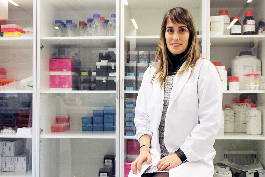
"Early diagnosis does not only prevent ulcers from developing, it can also prevent the development of cancer", says PhD student Silvia Fontenete from the Nucleic Acid Center at the University of Southern Denmark/Laboratory for Process Engineering, Environment, Biotechnology and Energy, University of Porto in Portugal. She is the lead author of an article on the research results in journals PLOS One.
Ulcers are often caused by the bacteria Helicobacter pylori, which produces ulcers in the stomach or duodenum. Usually the doctor retrieves a tissue sample from the stomach and has it analyzed. You can also take a breath test, where molecules in the patient's breath are analyzed, but this technique is not always reliable, says Silvia Fontenete.
Scientists have for some time been able to make tissue samples from the stomach glow fluorescent green if the tissue is infected with H. pylori bacteria. For this the scientist will need to retrieve a piece of the stomach for analysis in the laboratory.
The researchers' new approach is to make the stomach glow bright fluorescent green WITHOUT taking tissue samples from it.
"Our laboratory experiments suggest that one day it will be possible for doctors to send some specially designed molecules down in the stomach, where they will make H. pylori glow brightly green ", explains Silvia Fontenete.
To see the green light the doctor will send a small micro-camera into the stomach.
The scientists report to have made H. pylori glow green in artificial tissue in the laboratory - this was tissue that mimics the lining of the human stomach.
"We believe that the same can happen in a real human stomach", says Silvia Fontenete.
To achieve their results, the researchers had to overcome two major challenges: The first challenge was to create the special molecules that can both detect H. pylori bacteria and function at approx. 37 degrees Celsius, which is the temperature of the human stomach. The second challenge was that the molecule should be able to function in the extremely acidic environment of the stomach.
Both challenges were solved by working with the so-called Locked Nucleic Acid (LNA), some special synthetic molecules developed by Silvia Fontenente's research director, Professor Jesper Wengel at University of Southern Denmark. LNA are artificial RNA-like molecules which can bind to microRNA molecules. They are extremely stable and can operate at lower temperatures and in more acidic environments than other molecules.
"We have been lucky: The first molecules I created turned out to do the job right away. I could have been more unfortunate, and it would have taken several years”, says Silvia Fontenete.
Contact Professor Jesper Wengel, Nucleic Acid Center, Department of Physics, Chemistry and Pharmacy and the Department of Biochemistry and Molecular Biology. Email: jwe@sdu.dk. Phone: + 45 65502510 and +45 20846872.
Photo of Silvia Fontenete: Birgitte Svennevig/SDU.
Ref: Fontenete, Sílvia; Guimarães, Nuno; Leite, Marina; Figueiredo, Céu; Wengel, Jesper; Azevedo, Nuno Filipe. Hybridization based detection of Helicobacter pylori at human body temperature using advanced locked nucleic acid (LNA) probes. PLoS ONE (2013), 8(11).